
The 3 Normal Age-Related Changes
Night vision depends on how much light reaches the retina and how efficiently the retina processes it in low-light conditions. Three separate structures degrade this pathway with age, and their effects compound:
1. Senile Miosis (Smaller Pupil)
The pupil's ability to dilate in darkness declines steadily with age. A 20-year-old's pupil can dilate significantly wider in low light than a 60-year-old's. Less pupil dilation means less light physically reaches the retina — by age 60, research estimates the retina receives roughly a third of the light it did at age 20 under identical dim conditions, purely from this mechanism.
2. Lens Yellowing and Light Scatter
The natural crystalline lens progressively yellows with age (a change distinct from cataract, though it's on the same continuum) and becomes less optically clear. This scatters incoming light rather than focusing it cleanly — which is precisely what produces the glare, halos, and starbursts many people notice around oncoming headlights at night, even before cataracts are diagnosed as visually significant.
3. Photoreceptor and Neural Changes
Rod photoreceptors — the cells responsible for low-light vision — decline in density and sensitivity with age, and the neural pathways that process dim-light signals slow down. The practical result is slower dark adaptation: it takes an older eye measurably longer to adjust after going from a lit environment (like a gas station) into darkness, a critical few seconds on the road.
Cataracts — the Most Common Treatable Cause
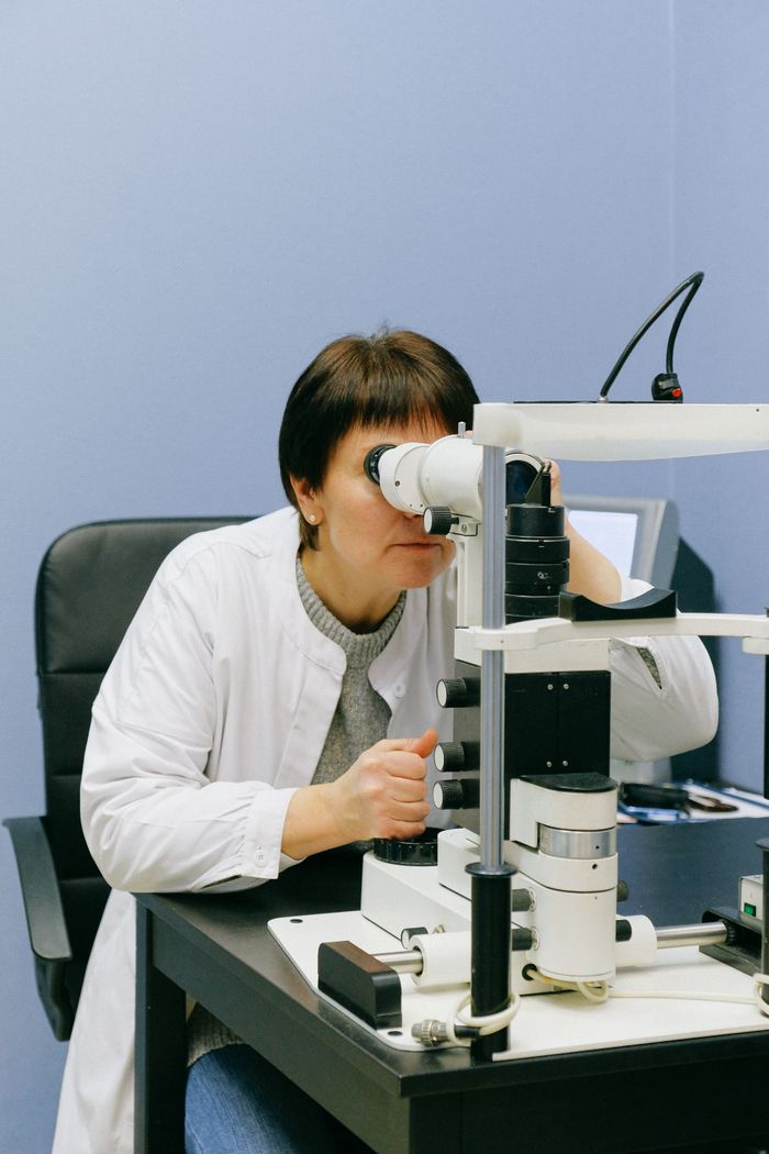
Cataracts — clouding of the natural lens — are extremely common with age and are frequently first noticed as a night-driving problem rather than a daytime one. Glare and halos around headlights can appear well before a cataract is advanced enough to reduce a standard daytime visual acuity score, because oncoming headlights create exactly the high-contrast, low-ambient-light scenario where lens clouding and light scatter are most noticeable.
This matters because, unlike most causes of night vision decline, cataracts have a direct, highly effective fix: surgical lens replacement. If glare sensitivity at night is your main complaint, a cataract evaluation is a reasonable, specific next step — this is not a "just live with it" category of aging change.
Other Disease-Driven Causes
| Condition | How it affects night vision |
|---|---|
| Early macular degeneration | Difficulty adapting to dim light and needing brighter light for near tasks are recognized early/intermediate AMD symptoms |
| Diabetic retinopathy | Retinal blood vessel damage can impair both central and low-light vision depending on severity and location |
| Vitamin A deficiency | Impairs rhodopsin regeneration in rods — the classic textbook cause of night blindness, rare in well-nourished populations |
| Retinitis pigmentosa | A rare inherited condition causing progressive rod photoreceptor loss, with night blindness often the first symptom, typically starting decades earlier than age-related decline |
When to See a Doctor
Gradual, symmetric (both-eyes) night vision decline over years, especially past age 50–60, is consistent with the normal aging changes described above. A comprehensive eye exam is still worthwhile to rule out treatable contributors, but it isn't necessarily urgent. What does warrant a prompter evaluation:
- Sudden change rather than gradual, over weeks rather than years
- One eye significantly worse than the other
- Accompanied by blurred central vision or wavy/distorted straight lines — possible signs of macular degeneration progression
- A family history of retinitis pigmentosa combined with early-onset night blindness
What Actually Helps
- Cataract evaluation: the highest-impact fix when lens clouding/glare is the driver — direct and effective.
- Anti-reflective lens coatings: for glasses-wearers, AR coatings reduce internal lens reflections that worsen glare from headlights.
- Avoid staring directly at oncoming headlights: a small habit shift — glancing slightly toward the right edge of the lane — reduces glare exposure and preserves dark adaptation.
- Clean windshields and headlights: a hazy windshield or clouded headlight lenses (common on older vehicles) scatter light in exactly the way an aging cornea/lens does, compounding the problem.
- Address underlying AMD or diabetic retinopathy with appropriate medical management if either is diagnosed as a contributing factor.
Zinc supports the vitamin A pathway rods depend on
Zinc is required to mobilize vitamin A from liver stores for use in the retina's rhodopsin cycle, which is why it's part of the original AREDS2 formula. VisiFlora and iGenics both include the AREDS2-dosed zinc and carotenoid stack as part of broader retinal support formulas — not a treatment for cataracts or AMD, but relevant nutritional support alongside proper medical evaluation.
The Nutrition Angle: Vitamin A and Zinc
Vitamin A's role in night vision is one of the most firmly established nutrient-vision links in all of nutrition science — vitamin A (as retinal) is a direct structural component of rhodopsin, the light-sensing pigment that must be regenerated after each exposure to light for rods to keep functioning in the dark. Severe vitamin A deficiency causing night blindness remains a leading cause of preventable vision impairment in regions with malnutrition, per WHO data.
For most adults in well-nourished countries, however, outright vitamin A deficiency is uncommon, and supplementing extra vitamin A when you're not deficient does not improve night vision beyond a normal baseline — more is not better once intake is adequate, and vitamin A has a narrower safety margin than most eye-health nutrients at high doses. The more broadly relevant nutrient here is zinc, which supports vitamin A transport and utilization and is part of the clinically-tested AREDS2 formula for macular health — a different and better-supported goal than "boosting" night vision beyond normal.