The 3 Stages of AMD
The current clinical staging system was established by Ferris et al. in 2013 (Ophthalmology) and adopted by the American Academy of Ophthalmology. AMD is classified by the size and number of drusen deposits and the presence of retinal pigment epithelium (RPE) changes, visible on fundus examination.
Stage 1: Early AMD
Defined by the presence of medium-sized drusen (63–124 µm in diameter) in one or both eyes, with no pigmentary abnormalities and no vision loss. Drusen are small yellow deposits of cellular debris that accumulate between the RPE and Bruch's membrane (the basement membrane beneath the retinal pigment epithelium). They are the hallmark finding of AMD.
At this stage, the photoreceptors and RPE cells are intact. There is no detectable change in visual acuity, contrast sensitivity, or other vision functions. The person has no awareness that anything is wrong. Early AMD is effectively invisible without a clinical examination.
Stage 2: Intermediate AMD
Characterized by one or more large drusen (≥125 µm — roughly the width of a retinal blood vessel), and/or pigmentary abnormalities in one or both eyes (areas of RPE hyperpigmentation or depigmentation). At this stage, some functional vision changes may begin — though they are often subtle enough to be attributed to aging or not noticed at all.
This is the stage where the AREDS2 formula was most studied and showed the greatest benefit: 25% reduced risk of progression to advanced AMD. Intermediate AMD is the critical intervention window.
Stage 3: Late AMD
Late AMD has two subtypes with different mechanisms:
- Geographic atrophy (GA) / dry AMD: Gradual, progressive degeneration and death of photoreceptors and RPE cells in the macula. Central vision loss expands slowly over months to years. Accounts for ~85–90% of AMD cases but ~10% of severe vision loss from AMD.
- Neovascular AMD / wet AMD: Abnormal blood vessels grow beneath the retina from the choroidal layer (choroidal neovascularization, CNV). These vessels leak fluid and blood rapidly, causing sudden and severe central vision loss. Accounts for ~10–15% of AMD cases but ~90% of AMD-related severe vision loss. Wet AMD can develop from dry AMD suddenly — which is why monitoring is critical.
Symptoms by Stage: What You Actually Notice
| Stage | What the doctor sees | What you notice |
|---|---|---|
| Early AMD | Medium drusen (63–124 µm), no pigment changes | Nothing — completely asymptomatic |
| Intermediate AMD | Large drusen (≥125 µm), RPE pigment changes | Mild blur, dim-light difficulty, needing brighter reading light |
| Late AMD (Atrophic) | Geographic atrophy — patches of RPE/photoreceptor loss | Growing central dark or blurred area, reduced color vibrancy |
| Late AMD (Neovascular) | Choroidal neovascularization, subretinal fluid/blood | Sudden vision loss, straight lines appear wavy or bent (metamorphopsia) |
Intermediate AMD symptoms to watch for in everyday life:
- Adaptation time in dim light: Taking longer than usual to adjust when moving from a bright to a dark environment (e.g., entering a dim restaurant)
- Needing more light: Previously comfortable reading light now seems insufficient
- Central blur: Reading fine print becomes more difficult even with correct glasses prescription
- Reduced contrast: Colors appear less vivid or washed out, particularly in lower light conditions
- Difficulty with faces: Recognizing faces at a distance becomes harder when looking directly at them (because central vision is affected; peripheral remains intact)
Why Early AMD Is Particularly Dangerous
The absence of symptoms in early AMD is its most dangerous feature. By the time someone notices visual changes, the disease has typically progressed to at least intermediate stage — and often beyond. The window for nutritional and preventive intervention is completely asymptomatic.
Additionally, AMD disproportionately affects central vision (the macula = central 5° of the visual field). The peripheral retina compensates well; people often unconsciously adapt, using peripheral vision to perform tasks that previously relied on central vision. This compensation can mask significant macular deterioration until the central scotoma becomes large enough to be undeniable.
The practical implication: regular dilated eye exams — not symptom monitoring — are the only reliable way to catch AMD at its most treatable stage. The American Academy of Ophthalmology recommends a baseline dilated exam at 40, and regular follow-up exams every 1–2 years from age 65 onwards (or sooner with risk factors).
The Amsler Grid: How to Screen at Home
The Amsler grid is the standard home monitoring tool for AMD, particularly for people who have already been diagnosed with intermediate AMD and are monitoring for signs of conversion to wet AMD. It consists of a grid of horizontal and vertical lines with a central fixation dot.
To perform the test correctly:
- Wear your reading glasses (if you use them)
- Hold the grid at normal reading distance (approximately 30 cm / 12 inches)
- Cover one eye and focus on the central dot with the other
- Note whether the surrounding lines appear straight, uniformly spaced, and complete — or whether some appear wavy, bent, or missing
- Repeat with the other eye
Any new distortion, waviness, or blank areas that weren't there before warrants a prompt call to an eye care professional — not waiting for your scheduled appointment.
Our free online vision screening includes an interactive Amsler grid as part of a 4-part eye health check. It can give you a baseline to compare against, but is not a substitute for a clinical dilated examination.
Important limitation: The Amsler grid detects intermediate-to-advanced AMD distortion. It will not detect early AMD, which is asymptomatic by definition. Do not use a normal Amsler grid result as reassurance that you don't have early AMD.
Try the interactive Amsler grid (+ 3 more vision tests)
Our free 5-minute vision screening includes visual acuity, color vision, astigmatism, and Amsler grid — results in under 5 minutes.
Who Is Most at Risk
Risk factors for AMD fall into two categories: non-modifiable and modifiable:
Non-Modifiable Risk Factors
- Age: The single strongest risk factor. AMD is rare before 50, present in ~10% of adults 66–74, and ~30% of adults 75–85 (NEI prevalence data). The 85+ age group has the highest risk.
- Family history: Having a first-degree relative with AMD approximately doubles risk. Specific genetic variants in complement factor H (CFH) and ARMS2 genes are strongly associated with AMD susceptibility.
- Race: AMD is significantly more prevalent in Caucasian and Chinese populations compared to African American populations. The reasons involve both genetic and potentially pigmentation-related (ocular protective pigment) factors.
- Sex: Women have a modestly higher prevalence than men, partly due to longer average lifespan.
Modifiable Risk Factors
- Smoking: The strongest modifiable risk factor. Current smokers have 2–4× the AMD risk of non-smokers. Risk remains elevated for years after quitting but decreases over time. (Mitchell P et al., 2002, Blue Mountains Eye Study)
- Cardiovascular disease / hypertension: Shares vascular pathophysiology with AMD (Bruch's membrane functions as a vascular filter). High blood pressure is independently associated with AMD risk.
- UV exposure: Cumulative sunlight exposure is a contributing factor, particularly blue-wavelength UV. Protective sunglasses (UV-blocking, wraparound) are recommended.
- Diet: Diets low in lutein, zeaxanthin, omega-3 fatty acids, and antioxidants are associated with higher AMD risk. The AREDS dietary component analysis showed that participants in the highest quintile of dietary lutein/zeaxanthin intake had significantly lower risk of AMD progression.
- Obesity: BMI >30 is associated with increased AMD risk and faster progression to advanced AMD.
When to See a Doctor (and What to Ask For)
See an eye care professional promptly if you notice any of the following:
- Straight lines appearing wavy, bent, or distorted (metamorphopsia)
- A blurry or dark area in the center of your vision (even if small)
- Colors appearing suddenly less vivid or washed out
- Any new or sudden change in vision quality — do not wait for a scheduled appointment
At a routine exam, ask specifically:
- "Can you look for drusen or any signs of macular degeneration?" — some routine eye exams focus primarily on refraction (glasses prescription) and don't automatically include a dilated fundus exam
- "Can you take fundus photos so we have a baseline to compare at future visits?"
- If you have risk factors: "Should I be on AREDS2 supplements?" and "How often should I come in for monitoring?"
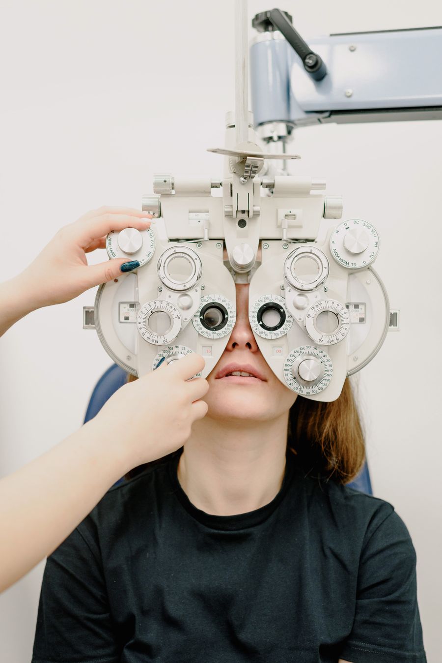
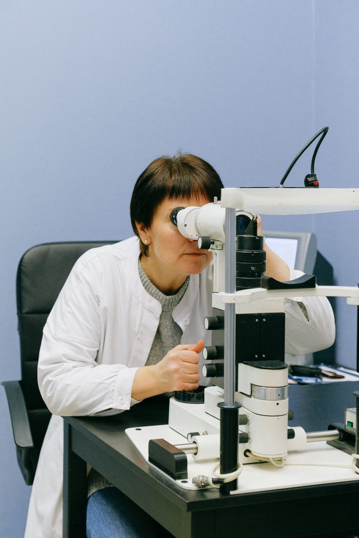
Dilated fundus examination (where eye drops widen the pupil) is required for accurate drusen assessment — standard non-dilated exams can miss early macular findings. Optical coherence tomography (OCT) imaging provides even more detailed cross-sectional imaging of retinal layers and is increasingly standard in AMD monitoring.
What You Can Do Nutritionally at Each Stage
Nutrition is the best-evidenced modifiable intervention for AMD — specifically the AREDS2 formula.
No AMD / Early AMD
For adults with no AMD or early AMD (small drusen only), the NEI does not currently recommend AREDS2 supplements — they were not tested and did not show benefit in this population in the AREDS2 trial. The recommendation for this group is dietary: maximize lutein and zeaxanthin from dark leafy greens, omega-3 fatty acids (fatty fish twice weekly), and antioxidant-rich vegetables. However, given the safety of lutein/zeaxanthin at 10–20 mg/day and the typical gap between dietary intake and protective amounts, many ophthalmologists support early supplementation as preventive strategy.
Intermediate AMD
This is the group where AREDS2 supplements have the strongest evidence. The NEI explicitly recommends AREDS2 formula for people with intermediate AMD in one or both eyes, or advanced AMD in one eye. The formula reduced progression risk by 25% over 5 years. → AREDS2 formula: complete breakdown
Supplements exceeding the AREDS2 carotenoid doses (such as 20 mg lutein vs. 10 mg AREDS2) have mechanistic rationale and MPOD evidence but have not been compared head-to-head to AREDS2 in large RCTs for AMD progression. → How much lutein per day?
Late AMD
For late AMD (geographic atrophy or neovascular AMD), the AREDS2 formula is still recommended to protect the fellow eye. Neovascular AMD is now primarily treated with anti-VEGF intravitreal injections (ranibizumab, aflibercept, bevacizumab) administered by a retinal specialist — supplements do not replace this treatment. Geographic atrophy currently has limited treatment options (though complement inhibitors are an emerging area).
VisiFlora and iGenics — AREDS2+ carotenoid formulas
VisiFlora: 20 mg lutein, 4 mg zeaxanthin, plus the full AREDS2 stack and gut-barrier nutrients. iGenics: 20 mg lutein, 10 mg zeaxanthin, plus bilberry 480 mg, saffron 20 mg, astaxanthin 4 mg, and turmeric with BioPerine. Both offer a money-back guarantee. Best for: intermediate AMD risk, adults 50+, screen workers.