What Floaters Actually Are
The inside of your eye, behind the lens, is filled with the vitreous humor — a clear, gel-like substance that helps the eye hold its round shape. In youth, the vitreous is a uniform, dense gel firmly attached to the retina. With age, it gradually liquefies and its collagen fibers clump together into small strands and particles.
Those clumps float within the more liquid vitreous and cast tiny shadows onto the retina as light passes through them — which is what you perceive as specks, threads, rings, or cobweb-like shapes drifting across your field of vision, especially noticeable against a bright, plain background like a clear sky or a white wall.
When Floaters Are Normal Aging
The process described above has a clinical name: posterior vitreous detachment (PVD) — the point at which the shrinking vitreous gel fully separates from the retinal surface it was attached to. PVD is extremely common with age; population studies estimate it affects a majority of people by their mid-60s to 70s, and it can occur even earlier in people who are nearsighted (myopic) or who have had cataract surgery.
A PVD itself is not a disease and does not damage vision. The floaters it produces are typically:
- Gradual in onset — appearing over days to weeks, not instantaneously
- Few in number — a handful of new specks or one or two larger strands, not a sudden "shower"
- Stable over time — the same floaters, in roughly the same pattern, rather than new ones continuing to appear
- Not accompanied by flashes of light or vision loss
In this typical pattern, floaters usually become progressively less bothersome over weeks to months — not because they disappear, but because they settle lower in the vitreous (out of the direct visual axis) and because the brain adapts to filter out a stable, unchanging visual artifact, a process called neuroadaptation.
Red-Flag Symptoms — See a Doctor Same-Day
A small percentage of PVD cases occur with enough force to tear the retina where the vitreous was most firmly attached. A retinal tear can progress to a retinal detachment — a sight-threatening emergency — within days if untreated. The warning signs are specific and worth memorizing:
Get evaluated the same day if you notice:
- • A sudden shower of many new floaters, appearing over minutes to hours rather than days
- • Flashes of light (photopsia) — brief streaks or sparks of light, especially in peripheral (side) vision, particularly in a dark room
- • A dark curtain, shadow, or veil moving across part of your visual field
- • Any sudden loss of peripheral or central vision
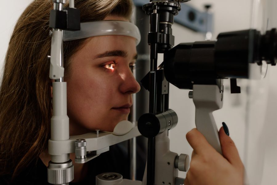
Any one of these on its own warrants a prompt exam; the combination of new floaters plus flashes plus a shadow is the classic presentation of an evolving retinal detachment. This is not a "wait and see if it gets better" situation — a dilated retinal exam is needed to rule it out, and if a tear is found, it can typically be sealed with laser photocoagulation or cryopexy in an outpatient procedure before it progresses to a full detachment.
Who's at Higher Risk
| Risk factor | Why it matters |
|---|---|
| Age 50+ | PVD becomes progressively more common with age as vitreous liquefaction advances |
| High myopia (nearsightedness) | A longer eyeball shape places more traction on the vitreous-retina attachment, increasing tear risk |
| Previous cataract surgery | Removing the natural lens can accelerate vitreous changes and PVD onset |
| Eye trauma | Blunt or penetrating injury can cause acute vitreous hemorrhage and floaters |
| Diabetic retinopathy | Abnormal retinal blood vessels can bleed into the vitreous, causing a sudden floater shower |
| Eye inflammation (uveitis) | Inflammatory cells and debris in the vitreous can appear as floaters |
Treatment Options
For typical, benign age-related floaters, the standard approach is observation — no treatment is medically necessary, and the large majority of people adapt to them within months. For floaters severe enough to meaningfully interfere with daily vision, two procedures exist:
- Laser vitreolysis: a laser breaks up large floaters into smaller, less noticeable fragments. Reserved for select cases; not all floaters are good candidates depending on their position and composition.
- Vitrectomy: surgical removal and replacement of the vitreous gel. Effective at eliminating floaters but carries surgical risks (retinal detachment, cataract formation, infection) that must be weighed against the relatively benign nature of most floaters.
Retinal tears and detachments are treated urgently and separately from routine floater management — typically with laser photocoagulation, cryopexy, or, for a full detachment, surgical repair (pneumatic retinopexy, scleral buckle, or vitrectomy depending on severity).
No supplement treats floaters — but overall retinal health still matters
Be skeptical of any product claiming to dissolve or eliminate existing floaters — no ingredient has clinical trial evidence for that. What a comprehensive formula can plausibly support is the broader antioxidant and microvascular environment of the retina over time. VisiFlora combines the AREDS2 carotenoid stack with gut-barrier nutrients; iGenics combines high-dose carotenoids with bilberry and saffron.
What Nutritional Support Can and Can't Do
To be direct: there is no supplement, vitamin, or nutrient with clinical trial evidence for shrinking, dissolving, or eliminating existing vitreous floaters. Anyone marketing a product that way is overstating what the science supports, and that kind of floater should be evaluated by an eye doctor, not "treated" with a pill.
What a well-designed formula can reasonably claim is supporting the general health of the structures involved — antioxidant status in the retina and its blood supply — which is a different and much narrower claim than "gets rid of floaters." If you're specifically bothered by floaters, the appropriate first step is always a dilated eye exam to confirm there's nothing else going on, not a supplement purchase.